Leg Bones Diagram / Skeletal System Diagrams - Your leg bones are the longest and strongest bones in your body.. A leg bone is a bone found in the leg. They allow you to move and provide support for your upper body. Other sets by this creator. The foot bones shown in this diagram are the talus, navicular, cuneiform, cuboid, metatarsals and calcaneus. Learn how to draw the femur, patella, tibia, and fibula in this lesson!
Diagram of blood and nerve supply to bone. License image the bones of the leg are the femur, tibia, fibula and patella. Other sets by this creator. Here are a few anatomical plates about the leg and the foot. The bones involved in it, however, are only the femur and the tibia, although the smaller bone of the leg, the fibula, is carried along in the movements of flexion, extension, and slight rotation that this joint.
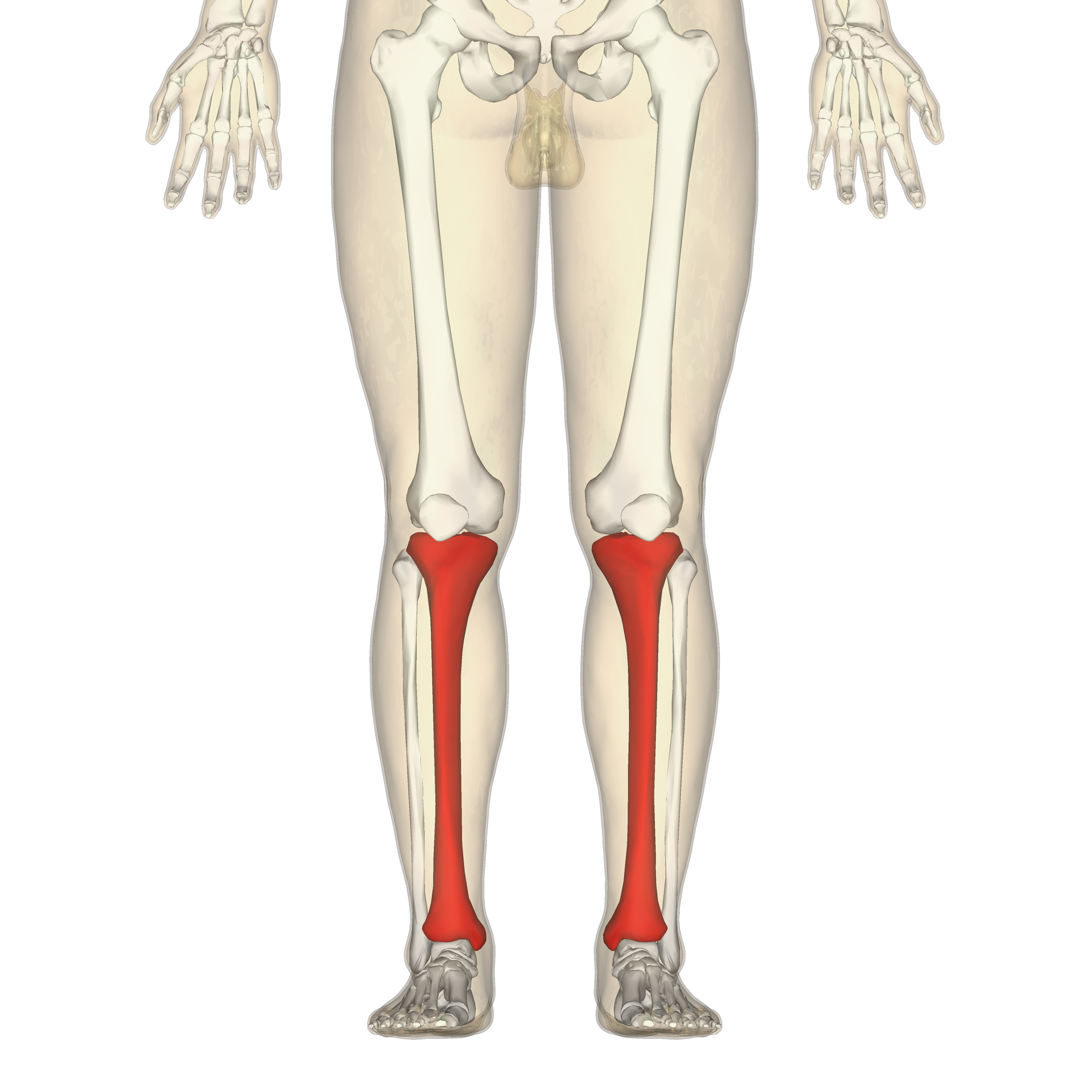
Diagram Knee Leg Bone Diagram Full Version Hd Quality Bone Diagram Wiringswitch Parrocchiecordenons It from 3.bp.blogspot.com The largest and most medial leg bone, forming both the knee and ankle joints. Quizzes on human skeletal system anatomy, bone anatomy, and bone markings. The femur in the thigh; Master leg and knee anatomy using our topic page. Click on the figures for a detailed view you will find the pelvic bones in the hip; These can include any the following: The tibia is the main bone of the leg, forming what is more commonly known as the shin. Your leg bones are the longest and strongest bones in your body.
The femur in the thigh;
The bones of the leg are the femur, tibia, fibula and patella. These simple labelled diagrams of the bones of the lower legs and feet and the bones of the arms and hands this diagram shows the skeletal structure of the leg (anterior view) and foot (dorsal view). Learn how to draw the femur, patella, tibia, and fibula in this lesson! The femur in the thigh; They allow you to move and provide support for your upper body. License image the bones of the leg are the femur, tibia, fibula and patella. Master leg and knee anatomy using our topic page. It expands at the proximal and distal ends, articulating at the knee and ankle joints respectively. Normal leg bones are relatively straight, but those affected by paget's disease are porous and figure 9. Pngtree offers bone diagram png and vector images, as well as transparant background bone diagram clipart images and psd files. When you stand or walk, all the weight of your upper body rests on them. These can include any the following: Bones in the human bodies and names.
Click on the figures for a detailed view you will find the pelvic bones in the hip; The largest and most medial leg bone, forming both the knee and ankle joints. Visit kenhub for more skeletal system quizzes. Human foot bones anatomy sketch of orthopedics medicine. The foot bones shown in this diagram are the talus, navicular, cuneiform, cuboid, metatarsals.
Leg And Knee Anatomy Bones Muscles Soft Tissues Kenhub from thumbor.kenhub.com Click now to learn more about the bones, muscles, and soft tissues tibia: Cheek bone (zygoma) upper jaw (maxilla). The patella in the knee; A leg bone is a bone found in the leg. The bones and joints in the feet experience wear and tear, so conditions that cause damage to the it is usually the result of a muscle imbalance when the long muscles of the lower leg overpower the. These can include any the following: Diagram of blood and nerve supply to bone. Learn how to draw the femur, patella, tibia, and fibula in this lesson!
Diagram of blood and nerve supply to bone.
Normal leg bones are relatively straight, but those affected by paget's disease are porous and figure 9. The bones involved in it, however, are only the femur and the tibia, although the smaller bone of the leg, the fibula, is carried along in the movements of flexion, extension, and slight rotation that this joint. The bones and joints in the feet experience wear and tear, so conditions that cause damage to the it is usually the result of a muscle imbalance when the long muscles of the lower leg overpower the. The foot bones shown in this diagram are the talus, navicular, cuneiform, cuboid, metatarsals and calcaneus. Your legs are two of your most important body parts. Click on the figures for a detailed view you will find the pelvic bones in the hip; Health diagram bone skeleton leg knee science anchor chart human human body. Click now to learn more about the bones, muscles, and soft tissues tibia: Blood vessels and nerves enter the bone. Most bones (particularly the long bones of the arms and legs — which make up the appendicular skeleton) have a hard outer shell known as cortical bone. Learn vocabulary, terms and more with flashcards, games and other study tools. A leg bone is a bone found in the leg. Quizzes on human skeletal system anatomy, bone anatomy, and bone markings.
Normal leg bones are relatively straight, but those affected by paget's disease are porous and figure 9. The foot bones shown in this diagram are the talus, navicular, cuneiform, cuboid, metatarsals and calcaneus. The bones and joints in the feet experience wear and tear, so conditions that cause damage to the it is usually the result of a muscle imbalance when the long muscles of the lower leg overpower the. Click now to learn more about the bones, muscles, and soft tissues tibia: Other sets by this creator.
Tibia Wikipedia from upload.wikimedia.org Click now to learn more about the bones, muscles, and soft tissues tibia: A leg bone is a bone found in the leg. Its lower end helps create the knee joint. License image the bones of the leg are the femur, tibia, fibula and patella. Each leg is made up of four bones. Click on the figures for a detailed view you will find the pelvic bones in the hip; Other sets by this creator. The foot bones shown in this diagram are the talus, navicular, cuneiform, cuboid, metatarsals and calcaneus.
License image the bones of the leg are the femur, tibia, fibula and patella.
Diagram and names of leg bones, diagram of foot and leg bones, diagram of leg bones, diagram of lower leg related posts of diagram of leg bones. The musculoskeletal segment of the leg, including the foot bones (ankle, heel bone, toe bones), fibula and tibia, knee, femur and femoral neck, hip and sacrum as well as the third, fourth. Lower jaw (mandible) collar bone. You'll learn about the muscles, bones, and other structures of each area of the leg. Pngtree offers bone diagram png and vector images, as well as transparant background bone diagram clipart images and psd files. Time to jump right into the biggest and strongest bones in the human body. These simple labelled diagrams of the bones of the lower legs and feet and the bones of the arms and hands this diagram shows the skeletal structure of the leg (anterior view) and foot (dorsal view). The foot bones shown in this diagram are the talus, navicular, cuneiform, cuboid, metatarsals and calcaneus. The foot bones shown in this diagram are the talus, navicular, cuneiform, cuboid, metatarsals and calcaneus. Its lower end helps create the knee joint. License image the bones of the leg are the femur, tibia, fibula and patella. The foot bones shown in this diagram are the talus, navicular, cuneiform, cuboid, metatarsals. Normal leg bones are relatively straight, but those affected by paget's disease are porous and figure 9.